Penny Rheingans and John Nichols
In Proceedings of Visualization '95 , IEEE Computer Society Press, pp. 393-396.
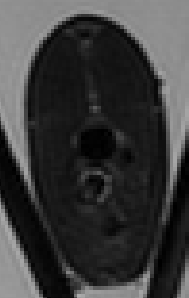
Figure 1. Raw MRI slice of rainbow trout. This image shows the second echo, which highlights the differences in water content. This is slice 43, located roughly two-thirds of the way from tail to head. The diagonal structures outside fish are the plexiglass support.

Figure 2. Segmented image (lice 43). Tissues and organs are colored to roughly correspond to their actual appearances: fat = off-white, kidney = dark brown, liver = light brown, intestine = yellow, and muscle = pink.
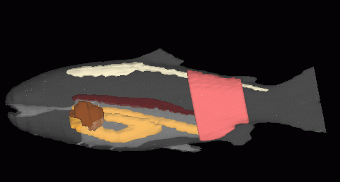
Figure 3. 3D trout colored by tissue type.
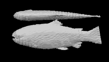
Figure 4. Fish exterior from segmented volume. Voxel artifact is visible in the block structure of the surface.
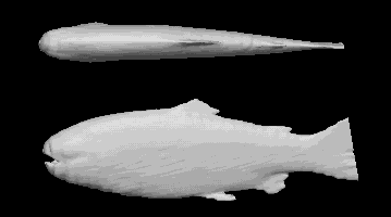
Figure 5. Fish exterior from blurred volume. Voxel artifact is greatly reduced, but fins have eroded somewhat.
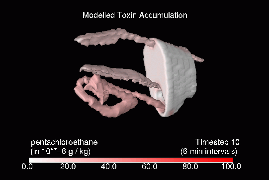
Figure 6. Tissues colored by concentration, after one hour of simulated exposure. Uptake happens most quickly in the GI tract.
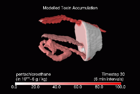
Figure 7. Tissues colored by concentration, after three hours of simulated exposure. Levels in tissue other than fat have begun to plateau, while concentrations in fat continue to climb.
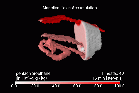
Figure 8. Tissues colored by concentration, after four hours of simulated exposure. Concentrations ain fat begin to dominate the toxin distribution.