Figure 1: Renal Artery Stenosis RAS


A region of renal artery where there is a build up in the thickness of the artery wall is called a stenosis as shown in figure 1. The stenotic region causes decreased pressure to the kidney leading to altered kidney function.

Figure 1: Renal Artery Stenosis RAS
Though conventional arteriography has been considered the
standard of reference in the diagnosis of renal artery stenoses(RAS), it is a
projectional technique that displays vessels through only a limited range of
viewing angle [1]. Spiral computed tomography(CT) was introduced
into clinical practice in 1989, and has been investigated and applied in a
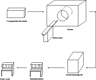
large number of studies [2]. A simple CT scanner is shown in figure
2.

Figure 2: Basic Components of a CT Scanner
Spiral CT can get a large volume of data in seconds, and offers more rapid examination time and lower radiation dose. Besides, the inherent contrast available with CT, along with reconstruction of cross sections, results in a good possibility to perform spiral CT angiography with excellent visualization of vessel lumina and stenoses from any viewing angle. Spiral CT angiography has the potential to become a less invasive screening tool [3]. The present study has focused on the accuracy of rendering techniques for evaluating renal artery stenoses.